
Ankle stiff squat depth suffers

Your heel rises. Your knee caves inward. Your lower back compensates.
Every squat, every lunge, every stair.
You’ve been told it’s your hips. Your glutes. Your thoracic mobility.
A 2024 randomised controlled trial found the ankle is often the actual restriction point — and that 8 weeks of a comprehensive corrective programme produced clinically significant improvements in dorsiflexion, balance, and proprioception.
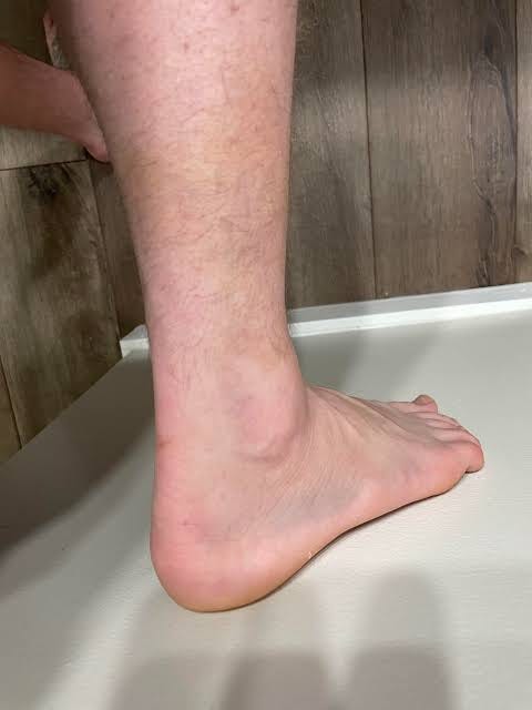
The restriction wasn’t in the muscles everyone was targeting. It was in the joint below them.
A 28-year-old software developer in Stockholm had been working on his squat for 14 months.
Hip mobility drills. Glute activation. Thoracic extension. The full corrective exercise stack.
His knee still caved on every heavy descent. His lower back still rounded at the bottom.
Nobody had measured his ankle dorsiflexion.
The weight-bearing lunge test would have shown him under 34 degrees — the threshold the 2024 study used to identify restricted ankles. Every squat he’d done for 14 months had been compensated from the ground up.
The ankle was the locked door. Everything else was people pushing on the walls.
If your squat mechanics break down consistently — heel rises, knee caves, or lower back rounds — and you’ve addressed hips, glutes, and thoracic mobility without lasting change, your ankle dorsiflexion may be the restriction point driving all three compensations simultaneously.
The ankle needs to dorsiflex adequately as the knee tracks forward over the foot during the squat descent. When it can’t — the heel rises, the tibia internally rotates, the knee follows inward, and the pelvis tilts forward to compensate.
Every rep reinforces the pattern.
A 2024 randomised controlled trial published in PLOS ONE tested a comprehensive corrective exercise programme on 30 female athletes with limited weight-bearing ankle dorsiflexion under 34 degrees.
After 8 weeks — the intervention group showed:
Dorsiflexion ROM: ES = 0.714 — a large clinically significant effect
Dynamic balance: ES = 0.423 — moderate significant effect
Proprioception: ES = 0.253 — significant improvement
Tibialis anterior and soleus muscle activity decreased during the overhead squat test — meaning the muscles worked more efficiently with less compensation after treatment.
Medial gastrocnemius activity in the descent phase decreased significantly — the calf stopped overworking once the joint could move correctly.
The control group showed no significant change across any measure.
[Source: Sohrabi T et al. Comprehensive corrective exercise program improves ankle function in female athletes with limited weight-bearing ankle dorsiflexion. PLoS One. 2024;19(10):e0312152. PMID: 39480882 → https://pubmed.ncbi.nlm.nih.gov/39480882/]
WHAT EVERYONE TRIES:
Glute activation / hip mobility drills / thoracic extension — while the ankle restriction continues loading every structure above it on every rep
WHY IT FAILS:
Limited ankle dorsiflexion changes lower limb biomechanics and neuromuscular control from the ground up. Working on structures above a mechanical restriction at the ankle is like tightening a rope that’s attached to a locked post. The structures above improve but the compensation pattern is maintained by the joint below.
WHAT WORKS:
Address the ankle first — soft tissue, joint mobility, range, strength — before layering squat volume and hip work on top of a restriction that’s driving the pattern
The Weight-Bearing Lunge Test
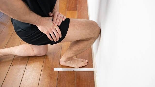
Before the protocol. A 30-second self-assessment.
Stand facing a wall. Place your foot 10–12cm from the wall. Lunge forward — try to touch your knee to the wall while keeping your heel flat on the floor.
If your heel rises before your knee reaches the wall — you have restricted ankle dorsiflexion.
Move the foot closer until the heel stays flat and the knee touches. Measure the distance. Under 9–10cm is generally considered restricted. Under 34 degrees on the formal lunge test is the threshold the study used.
This tells you more about why your squat breaks down than any video analysis of your hips.
The Chain Above a Restricted Ankle
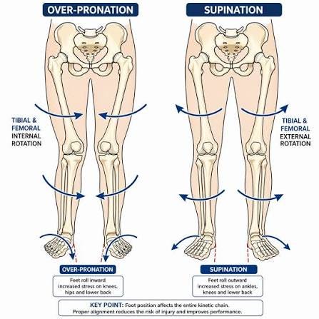
When the ankle can’t dorsiflex — the talus can’t glide backward to allow the shin to move forward over the foot.
The body finds the range somewhere else. The foot pronates — arch collapses, tibia internally rotates. The knee follows inward — valgus stress increases. The hip externally rotates to compensate for the tibial rotation below. The pelvis tilts forward to compensate for the hip above.
The lower back rounds at the bottom of the squat.
Every structure in that chain is responding correctly to the restriction. None of them are the actual problem. The ankle is.
This is why isolated glute work improves glute strength without changing squat mechanics. The glutes are fine. The ankle is locked. The chain above it adapts regardless of how strong the individual links are.
The study’s programme used four components in sequence: soft tissue mobilisation, joint mobilisation, stretching, then strengthening. The order matters — releasing the tissue before mobilising the joint, mobilising the joint before loading the range.
This is where the programme’s logic connects to recovery more broadly. Soft tissue quality — how the calf fascia and joint capsule respond to mobilisation — depends partly on systemic inflammation and recovery capacity. I can’t cover the nutrition and gut side of that here, but Coach Dylan wrote a piece specifically on how gut bacteria may drive systemic inflammation that affects tissue quality and recovery — including what a 2021 Stanford trial found about fermented foods versus fibre. It’s called You’re not fat. Your gut is starving if the recovery angle matters to you.
Which of these matches your situation:
→ Heel rises during squats — can’t keep it flat under load
→ Knee caves inward — worse on the dominant side
→ Lower back rounds at the bottom of the squat or lunge
→ Ankle clicks or feels restricted during dorsiflexion movements
→ Previous ankle sprain — mechanics never fully returned
Reply with whichever one matches.
Here’s the exact 4-component ankle corrective programme — soft tissue, joint mobilisation, stretching, and strengthening — in the order the RCT confirmed produces large effect size improvements in dorsiflexion within 8 weeks.
The Ankle Dorsiflexion Reset:
Release → Mobilise → Stretch → Strengthen
The study used four components in a specific sequence. The sequence is not arbitrary.
Release tight tissue before mobilising the joint. Mobilise the joint before loading the new range with stretching. Load the range with stretching before strengthening into it.
Reverse the order and each step is working against the restriction the previous step failed to address.
Soft Tissue Mobilisation — Calf and Plantar Fascia
(5 minutes — daily, always first)
The calf complex — gastrocnemius, soleus, and the posterior deep compartment — limits ankle dorsiflexion when the fascial tissue is restricted. The study included soft tissue mobilisation as the first component before any joint work.
Foam roller — calf:
Slow passes from Achilles insertion to the back of the knee. 2 minutes. Pause on tender areas for 10 seconds each. The gastrocnemius has two heads — roll the middle and lateral border separately.
Trigger point ball — soleus:
The soleus sits beneath the gastrocnemius. Cross one leg over the other at the knee to get bodyweight into the deeper muscle. 2 minutes. The soleus is the more clinically significant ankle dorsiflexion restrictor in desk workers — most foam rolling only reaches the gastrocnemius.
Why the soleus specifically: The study found medial gastrocnemius activity decreased significantly after treatment. But the soleus is the ankle plantar flexor that limits the tibia moving forward in the weight-bearing lunge position — the test used in the study. Address the soleus before the gastrocnemius.
